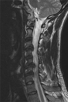
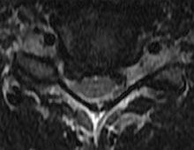
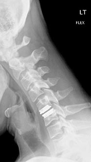
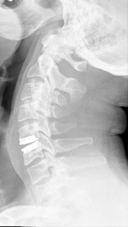
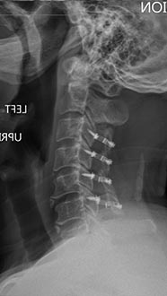
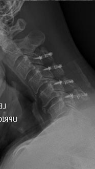
Aneterior Cervical Disc Replacement
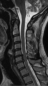
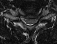
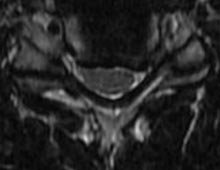
35 year old man presenting with severe neck pain with radiation down the right arm with right bicep weakness. MRI with large right C5-6 disc extrusion.
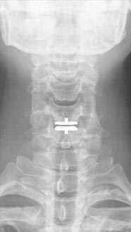
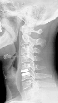
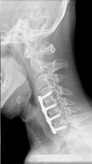
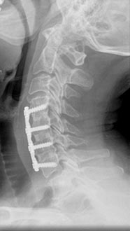
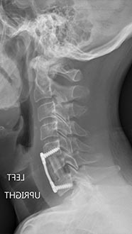
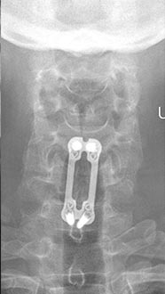
Postoperative xrays after C5-6 anterior cervical disc replacement. Complete resolution of arm pain and full painless range of motion of his neck.
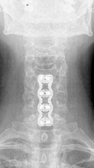
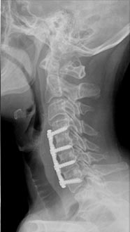
Aneterior Cervical Corpectomy
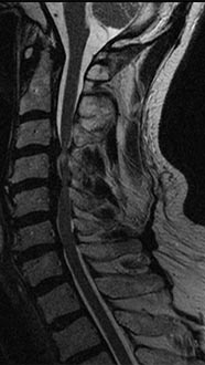
50 year old man presenting with left biceps and triceps weakness with numbness in his hand.
C6 corpectomy with complete resolution of the weakness and numbness.
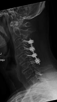
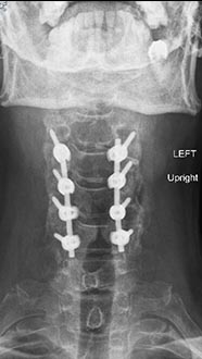
Posterior Cervical Laminectomy and Fusion
70 year old presented with spinal cord injury and quadriplegia after a fall.
Patient underwent laminectomy and fusion with complete resolution of the weakness and numbness.
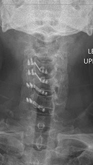
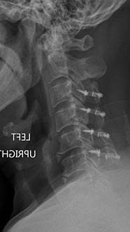
Cervical Laminoplasty
57 year old man presenting balance problems, constant numbness of both hands, and loss of dexterity in his hands with minor neck pain who had severe cervical stenosis. Cervical laminoplasty from C3 to C6 with motion preservation.
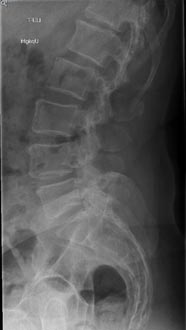
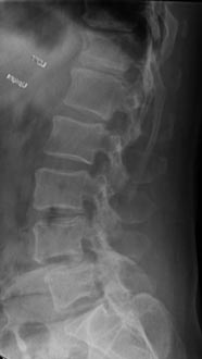
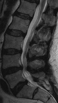
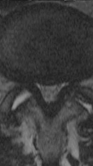
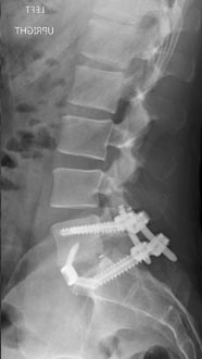
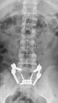
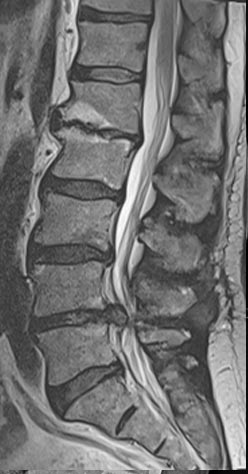
58 year old presenting with low back pain and bilateral leg pain especially when he stands for a few minutes or walks more than a few blocks.
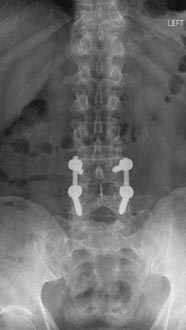
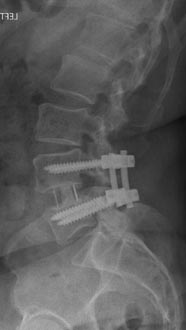
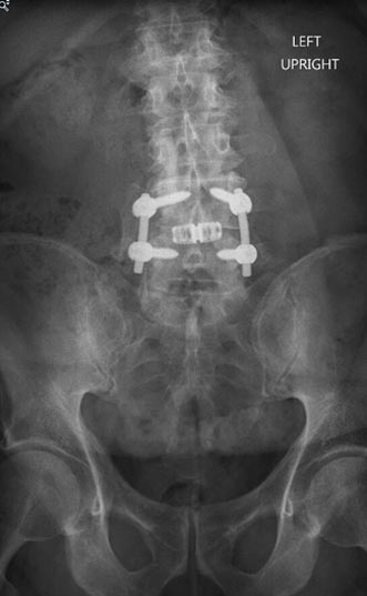
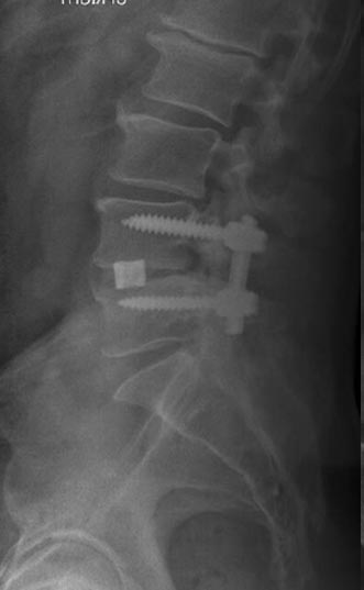
Solid fusion 2 years postop
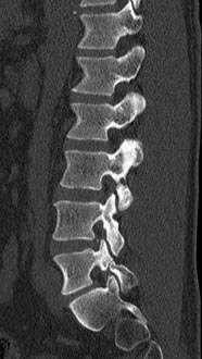
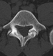
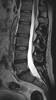
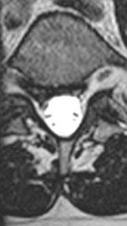
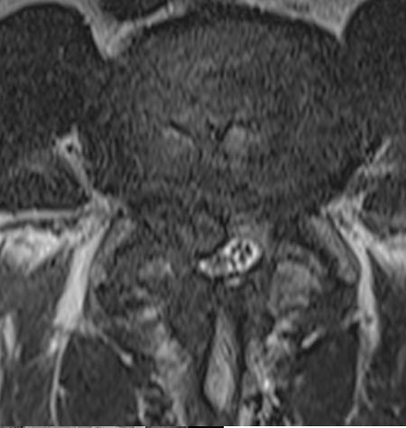
44 year old man with severe low back pain with intermittent radiation of the pain down the right leg. After undergoing conservative treatment, continued to have debilitating pain. MRI revealed a right paracentral disc herniation but CT scan revealed a unilateral pars fracture. After debating on microdiscectomy vs fusion, patient elected to undergo lumbar fusion. 4 months postop patient returned to surfing again. 9 months postop patient wrote “My recovery continues to do very well and we just got back from 8 days camping which including mountain biking, hiking, and a lot of work. Surfing almost every day and life is good…you have helped get my life and my families life back to normal.”
After procedure.
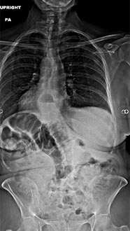
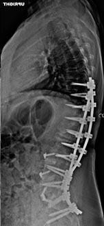
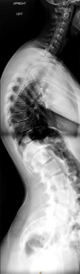
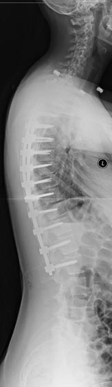
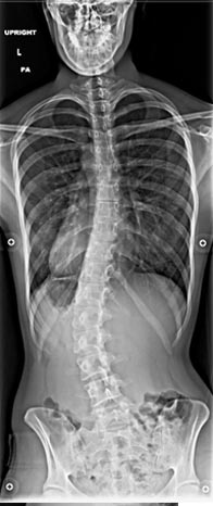
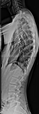
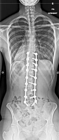
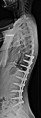
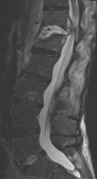
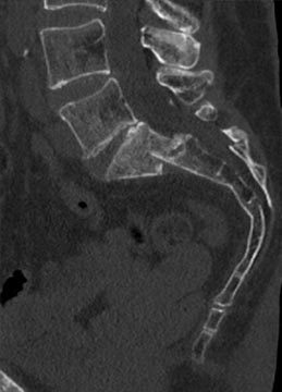
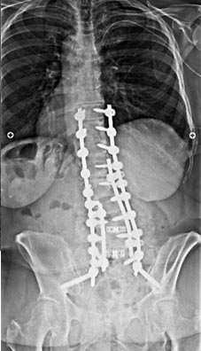
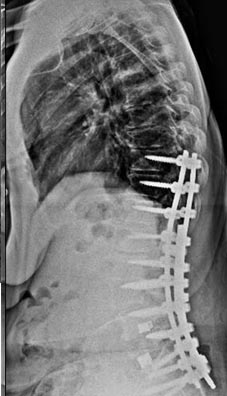
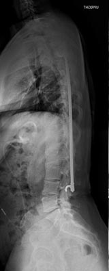
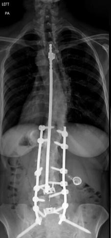
59 year old woman with Scholiosis with severe back pain.
1 year postop from surgery and pain free.
21 year old woman with very ridged 90 degree Scheuremann's kyphosis corrected to 40 degrees.
20 year old with severe low back pain from untreated idiopathic adolescent scholiosis who failed all conservative treatment with documented progression of scoliosis despite being done growing.
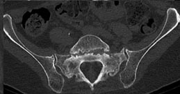
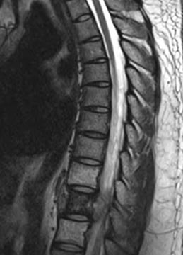
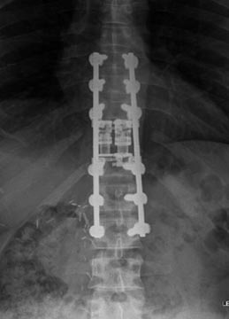
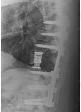
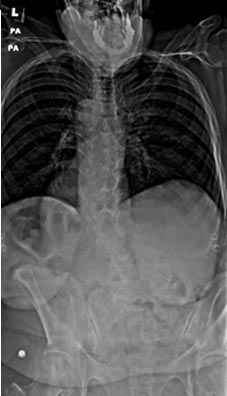
64 year old man with prostatitis who developed osteomyelitis of T12/L1 treated with an all posterior approach thoracolumbar fusion and debridement with antibiotic cement.
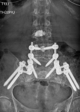
73 year old woman with osteoporosis who fell and fractured her pelvis. She was unable to walk and developed lumbopelvic dissociation (seperation of the spine from the pelvis and lower half of the body) from an unstable sacral fracture and displacement.
Lumbopelvic fixation with prophylactic cement augmentation and patient was immediately able to walk again.
41 year old male with metastatic renal cell carcinoma to the spine presenting with cord compression
Painfree after corpectomy and decompression of the spinal cord.
72 year old female who previously had a fusion with another surgeon who developed an infection and underwent multiple surgeries presenting with severe back pain with inability to stand upright.
46 year old woman who previously had Harrington rod spinal fusion from T4 to L3 for idiopathic adolescent scoliosis who presents with back pain and left leg weakness from severe lumbar stenosis distal to her previous fusion.
3 months postop, patient is pain free and weakness completely resolved. She is able to stand upright without pain and improved posture.
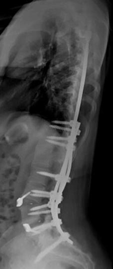
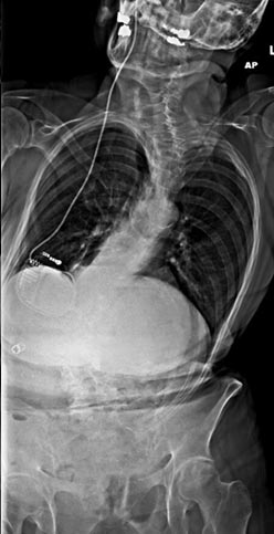
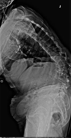
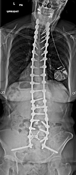
67 year old woman with Parkininsons presenting with neuromuscular kyphoscoliosis with inability to stand and look upright. Patient expressed that she was tired of talking to other people’s belly buttons.
T2 to sacrum/pelvis spinal fusion. Patient is pain free and able to stand upright and walk normally for the first time in years.
61 year old man with previous history of microdiscectomy at L4-5 presenting with severe back pain with radiating pain down the right leg with weakness. He had a massive disc extrusion on the right side. Options included revision discectomy vs. L4-5 revision decompression and fusion with transforaminal lumbar interbody fusion (TLIF). Patient did not want to go through the pains of recurrent disc herniation again and elected for the fusion.